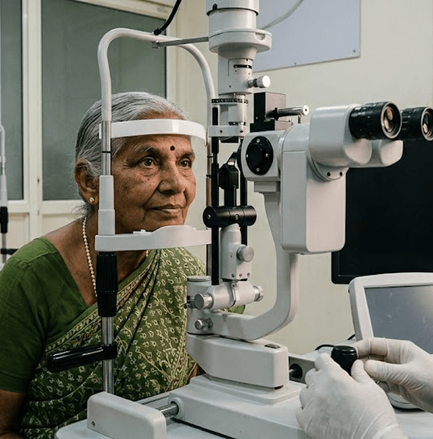






SPECIALITIES

Retina

Care for the light-sensing layer at the back of the eye, from diabetic eye disease to retinal detachment.

*Monday to Saturday, 9 am to 8 pm | Consultations from 8:45 am

Consultations

Habsiguda and Chanda Nagar. Laser, injections and surgery at Habsiguda.

Insurance and Schemes

EHS, CGHS and cashless insurance accepted. Aarogyasri covers selected retina procedures. WET AMD is only covered under ARS & EHS Anti vegif.

Treatment Type

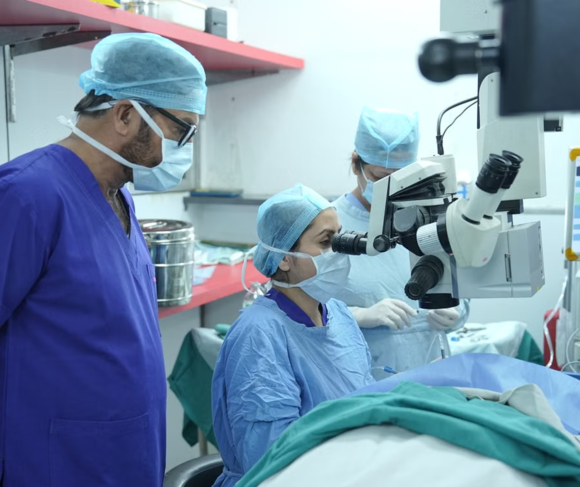
Laser and injections are outpatient procedures. Surgery is day care or a short stay.

Understanding Retina

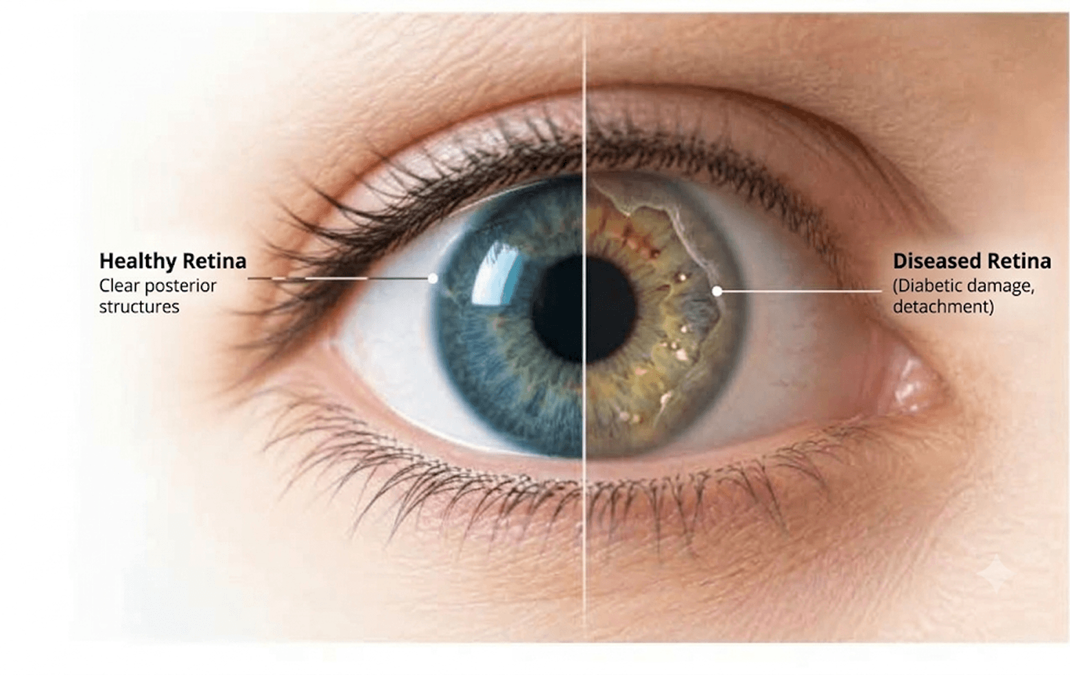
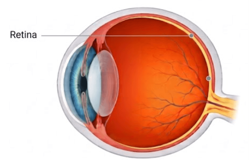
Healthy Retina
(Intact)
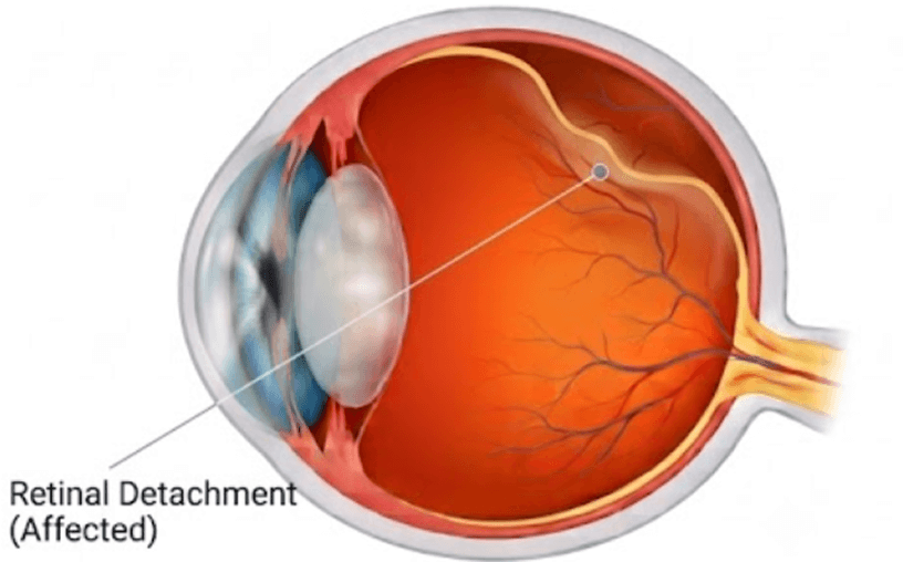
Affected Retina
(Distortion)

What happens to the retina?

The retina is a thin layer of light-sensitive tissue at the back of the eye. It turns light into signals that travel to the brain. The macula, at its centre, gives sharp central vision. The vitreous is the clear gel in front of the retina.

Retinal problems often cause no pain, and the damage can be permanent. Early detection protects vision.

  • Blurred or distorted vision, with straight lines looking wavy
  • A dark or blank patch in the centre of vision
  • A sudden shower of new floaters
  • Flashes of light
  • A curtain or shadow over part of your vision
  • Difficulty seeing in dim light
  • Colour & Contrast Issues

Causes: diabetes damaging the small blood vessels of the retina; ageing changes in the macula; vitreous gel pulling on the retina and causing tears; blocked retinal blood vessels; high myopia, which thins the retina; eye injury; premature birth; inherited conditions.

Risk factors: diabetes, especially long-standing or poorly controlled; high blood pressure and high cholesterol; age above 50; high myopia; family history of retinal disease; earlier retinal detachment or eye surgery; smoking.

  • Diabetic retinopathy: diabetes damaging the retina’s blood vessels
  • Retinal tear and detachment: the retina splits or peels away from the back of the eye
  • Age-related macular degeneration (AMD): wear of the central retina
  • Retinal vein or artery blockage
  • Macular hole and macular membrane
  • Vitreous haemorrhage and floaters: bleeding or debris in the gel
  • Retinopathy of prematurity (ROP): in premature babies (see Pediatric Ophthalmology)
  • Inherited retinal diseases
  • Keep blood sugar, blood pressure and cholesterol under control
  • If you have diabetes, get a dilated eye examination every year, or as advised (important)
  • Do not smoke
  • If you have high myopia, get your retina checked regularly (important)
  • Do not wait on sudden floaters, flashes or a shadow in your vision (important)

See a doctor the same day for new floaters with flashes, a shadow across your vision, or sudden vision loss. Early diabetic retinopathy often has no symptoms.

1. Diagnosis

A retinal problem is confirmed with detailed eye examination. Tests may include:

Dilated Retina Examination

Checks the whole retina

OCT

A cross-section scan of the macula

Retinal Photography

Records the retina to compare over time

2. Treatment

Early stages are watched while the underlying disease is controlled. Treatment is added when the retina is at risk.

When is treatment needed?

When there is a tear, detachment, leakage or swelling in the macula, or abnormal blood vessels. Retinal tears and detachments need treatment quickly.

Without surgery: 

Controlling blood sugar, blood pressure and cholesterol, with regular monitoring for early disease.

Treatment options:
  • Laser: Seals retinal tears and treats diabetic retinopathy. A short procedure under numbing drops.
  • Injections: Medicine injected into the eye for diabetic macular swelling, AMD and vein blockage. Usually a series of visits.
Surgical Options:
Retinal detachment repair: A scleral buckle, or vitrectomy with gas or silicone oil.

Macular hole surgery: Replaces cloudy or tugging gel with a temporary gas bubble to seal the center of your retina and restore central vision.

Vitrectomy: Removes blood, scar tissue or gel from inside the eye.

Got a diagnosis? Ensure you’re on the right track with a second opinion:

3. Recovery and Post-Op Care

First Week
  • Use eye drops exactly as prescribed
  • Keep the head position your doctor advises, sometimes face-down for days
  • With a gas bubble, do not fly or go to high altitude until your doctor confirms it has cleared. Tell any anaesthetist
  • Do not rub or press the eye
  • Wear sunglasses outdoors
Weeks 2-4
  • Avoid heavy lifting and strenuous exercise
  • Silicone oil, if used, is removed in a later surgery
  • Reading, TV and light activity are fine
Follow-up
  • Call us or visit immediately for increasing pain or redness, a drop in vision, new flashes or floaters, or a shadow across your vision.

After laser or an injection: glare or mild irritation for a day. Most people resume routine the next day. Use drops if prescribed.

Got questions about your recovery?

Planning Your Care

Comprehensive care, personalized for you. Know what is covered, what it costs and what to bring.

1.
Diagnosis
Diagnosis
2.
Treatment
Treatment
3.
Recovery
Recovery

Insurance & Schemes

What will it cost?
Consultation fee:

₹700
₹350 for EHS/CGHS beneficiaries

Treatment: Depends on the condition and treatment. Injections are a course of visits, so an estimate is given for the course

Before your visit
Bring:

Plan for: 1-2 hours for detailed examination

Unsure where to start? We’re here to help!

Frequently Asked Questions

Does diabetes always damage the eyes?

No. The risk rises with the years of diabetes and with poor control. Control and a yearly dilated check protect vision.

Early diabetic retinopathy has no symptoms. Dilation lets the doctor see the whole retina.

Most are harmless. New floaters with flashes of light need a same-day check, since they can signal a tear.

The eye is numbed first. Most people feel brief pressure.

It varies. Many people need a series. Your doctor reviews the response at each visit.

Usually yes, with surgery. The earlier it is treated, the better the chance of keeping vision.

No, not until your doctor confirms the gas has cleared.

Treatment can control the disease and prevent or reduce further vision loss. Options include laser treatment, injections into the eye, and surgery in advanced cases. Some patients require repeated treatment and long-term follow-up.

These are intravitreal injections, commonly anti-VEGF medicines, used to treat diabetic macular edema and certain complications of advanced diabetic retinopathy. Multiple injections may be needed.

Optical coherence tomography (OCT) is a painless, non-invasive imaging test that produces detailed cross-sectional images of the retina. It helps detect retinal swelling, fluid, and other structural abnormalities.

Usually, the injection is given under topical anesthetic eye drops.

No, there will not be any eye patch post procedure.

Diabetic, or
noticing floaters?

Book a retina consultation at Habsiguda or Chanda Nagar.

Scroll to Top